Anesthesia

Critical Care Anesthesia

Keeping patients comfortable, without pain and distress, and continuously monitoring vital signs is what makes this profession so important. 

What is Anesthesia?

Anesthesia is a medical treatment that prevents the feeling or sensation of pain with or without loss of consciousness during medical procedures. Anesthesiology also includes traditional intraoperative care and sub-specialties like chronic pain management and critical care.

Anesthesiologists are specialized doctors who administer anesthetics to patients, depending on the patient and the pain relief required. The various anesthetics administration routes include injection, inhalation, topical lotions, sprays, skin patches, and eye drops. Anesthesia is usually administered to patients who undergo complicated procedures like surgeries. 

What are Anesthetics?

Anesthetics are drugs that reduce or prevent pain. There are mainly three types of anesthetics used.

In a restricted area of the body, local anesthetics are drugs that, upon topical application or local injection, cause a reversible loss of sensory perception, especially pain. 

Regional anesthetics are local anesthetics that block the sensation perception from a larger body area, such as an arm, leg, or abdomen. 

General anesthetics are drugs that cause a reversible loss of consciousness, immobility, muscle relaxation, and sensation, especially pain. 

When local anesthetics or regional anesthetics are administered, the patient remains conscious, but during the administration of general anesthetics, the patient is unconscious and unaware of his/her surroundings. Anesthetics given to patients in critical care units are usually general anesthetics. 

What do you mean by critical care?

Critical care or Intensive care is a specialized type of care administered to patients with life-threatening injuries and illnesses. They need constant monitoring and comprehensive care and are usually admitted to intensive care units (ICU).

Health-care providers use a lot of different equipment like catheters, dialysis machines, feeding tubes, etc. Devices also monitor the patient’s vital signs and display them on monitors. These machines help keep patients alive, but they can also increase the risk of infection. 

Critical care anesthesia

Critical care anesthesia is a specific type of anesthesia where specialized anesthesiologists care for patients who have recently undergone major surgery or suffer from the effects of severe infections or trauma. 

Critical care anesthesiologists possess the medical knowledge and technical expertise to deal with emergencies. They work in intensive care units as essential doctors of care, also known as intensivists. They have the training to deal with cardiac and pulmonary resuscitation, airway management, advanced life support, and airway control; stabilizing the vitals.

They know how to stabilize and prepare patients for emergency surgeries. They coordinate and work closely with other specialists like surgeons to manage treatments for patients and deliver a full range of care. They act as a connecting link between surgeons and physicians to ensure that they work together effectively and for the same purpose. 

Critical care anesthesiologists continue to care and check on patients multiple times throughout the day and even at night. 

Some of the standard protocols critical care anesthesiologists follow are –

-Monitor the electrical activity of the heart

-Monitor the blood oxygen saturation

-Monitor vital signs like blood pressure, respiratory rate, and heart rate.

Usually, patients in the ICU are given intravenous saline so that they stay hydrated. 

During the coronavirus pandemic, the importance of critical care anesthesia has risen up. Anesthesiologists are working tirelessly to provide the necessary care required for an excessive amount of patients.

The management of COVID-19 patients includes specialized anesthetic care for patients with suspected and confirmed COVID-19, intubation outside operation theatres, oxygenation and ventilation support for acute hypoxemic respiratory failure, emergency management and participation in other patients care aspects.

Anesthesiologists have to be very careful as they will be handling most of the patients with respiratory disorders. Some requirements that need to be followed are 

-All suspected patients must be kept in isolation rooms before their procedure. 

-Anesthesiologists must wear Personal Protective Equipment at all times. 

-Operations, when conducted, should be done in a negative pressure or positive pressure isolation rooms. 

Effect on patients

Effect on patients

Critical care can have a significant impact on patients. Before being administered with anesthesia, patients must undergo a diagnostic assessment to determine his/her ability to survive the stress of anesthesia and surgery. 

Some difficult decisions must be made if the patient is critically ill or closer to death. Some patients may have to be resuscitated. Others can live but only with the aid of machines. The outcomes of these decisions must ensure that the patient undergoes minimal suffering. The entire team of healthcare workers and the patient’s family are involved in making these decisions. 

Anesthesiologists around the world work long hours and fatigable work shifts. Critical care anesthetists have more stress while handling multiple patients and continuously monitoring their vital signs. The coronavirus situation has heavily impacted the work-life of these anesthesiologists, causing even more challenges. 

Anesthesiologists are like the heroes behind a mask who ensure the patient’s safety while other specialists carry out their procedures. We have to thank them to keep us comfortable, pain-free, and in good health.

Do check our recent blog – https://www.saideephospital.com/2020/12/19/multiple-sclerosis/

Stay tuned for regular health updates – https://www.instagram.com/saideephealthcareofficial/

Multiple Sclerosis

Multiple Sclerosis – Neurology

An autoimmune disease is a condition where the immune system starts attacking its own body’s cells. This detrimental effect can lead to various complications and other diseases. The immune system cannot differentiate between foreign cells and its own cells, which leads to this condition. They can either target one organ, in Type 1 diabetes, or target multiple organs, in systemic lupus erythematosus. 

What is Multiple Sclerosis?

Multiple Sclerosis, also known as encephalomyelitis disseminate, is an autoimmune disease that attacks the myelin sheath of the nerve cells in the brain and spine. The myelin sheath is an insulating layer of proteins and fats that protects the axon of the nerve cells. This sheath allows proper electrical impulses to take place quickly and efficiently. It is made by two cells, oligodendrocytes, and Schwann cells.

During multiple sclerosis, the immune system starts eating away at the myelin sheath. It disrupts the electrical signals to pass effectively from cell-to-cell and causes a range of symptoms, from muscle weakness to paralysis in the legs. It can lead to permanent damage or deterioration of nerve cells. When a nerve fiber is exposed, the impulses can be slower or can stop completely.

Statistics

6.4% of women and 2.7% of men are likely to develop some kind of an autoimmune disease at some point in their lifetime. More than 2.3 million people in the world have multiple sclerosis. In India, almost every 5-10 per 1,00,000 individuals have multiple sclerosis. A study in 2015 stated that the number of people diagnosed with multiple sclerosis per year had almost doubled. 

Causes Multiple Sclerosis

Since it is an autoimmune disease, the exact cause of multiple sclerosis is unknown. However, a couple of factors can increase the risk of this disease. 

  • Age: multiple sclerosis is one of the most common disabilities in younger adults. It can develop at any age but is usually seen in adults from the age of 20-40. 
  • Sex: Women have a double or triple higher chance of developing multiple sclerosis than men. This could be due to the role of female hormones. 
  • Inheritance: multiple sclerosis is not hereditary but it can get passed down generations. The risk of siblings or children developing multiple sclerosis is higher. 
  • Infections: a research article conducted in 2014 states that multiple sclerosis can be linked with microbial infections like Chlamydia pneumonia and staphylococcus aureus produced enterotoxins.
  • Race: Caucasian people, usually of European descent, have the greatest risk of developing multiple sclerosis, whereas people of Asian or African descent, have the least risk.
  • Vitamin D: People with lower levels of vitamin D have a higher risk of developing multiple sclerosis.
  • Climate: climate does not cause this disease but it can worsen the symptoms of multiple sclerosis. This is usually temporary. 

Symptoms of Multiple Sclerosis

The symptoms of multiple sclerosis are unpredictable and can vary from person to person. Some symptoms of multiple sclerosis include:

  • Muscle stiffness, pain and spasms of the extremities.
  • Tingling and numbness in various parts of the body.
  • Vision problems like double vision, blurred vision and sometimes even loss of vision. 
  • Fatigue and weakness due to nerve deterioration in the spinal cord.   
  • Dizziness and incoordination in balance, often similar to vertigo.
  • Bladder, bowel and sexual dysfunction where patients lose bladder and bowel control. 
  • Cognitive issues like disorientation, shortened attention span and loss in memory. 
  • Emotional health is affected leading to irritability and mood swings.

Diagnosis: 

There is no specific test to diagnose multiple sclerosis. However, various other tests can rule out different diseases with similar symptoms to arrive at a conclusion. They are:

  1. Blood test: A blood test can check for specific biomarkers that may be associated with multiple sclerosis.
  2. Spinal tap: A puncture in the lumbar region of the spine can sample the cerebrospinal fluid for any abnormalities in antibodies. 
  3. Magnetic resonance imaging: An MRI can detect lesions on the brain and spinal cord which could be an underlying cause of multiple sclerosis. 
  4. Evoked potential test: An evoked potential test measures the speed of nerve messages along sensory nerves to the brain and a visual or electrical stimuli test used in the diagnosis of multiple sclerosis.   

Treatment: 

Treatment of Multiple Sclerosis

There is no cure for Multiple sclerosis. Treatment is focused on 3 main concerns:

  1. Treatment for attacks

Corticosteroids: they decrease inflammation but have various side effects like fluid retention, mood swings, and an increase in blood pressure and blood glucose levels.

Plasma exchange: exchanging plasma in the blood for albumin is used when the patient doesn’t respond to steroids

  1. Treatment to slow the progression

Treatment in the early stages can lower the relapse rate, slow the formation of new lesions, and reduce the risk of brain atrophy. For primary-progressive MS, ocrelizumab (Ocrevus) is the only FDA-approved disease-modifying therapy. There are various options available for relapses of MS such as Injectables (Interferon beta medication, Glatiamer acetate), oral medication (Dimethyl fumarate, Teriflunomide), or infusion treatments (Natalizumab). 

  1. Treatment for symptoms

Physiotherapy: Stretching and strengthening exercises can reduce muscle weakness and improve gait problems. 

Muscle relaxants: Relieves muscle stiffness and spasms.

Medications to reduce fatigue and improve mood: Selective serotonin reuptake inhibitors can be used for depression and Amantadine can decrease fatigue. 

Conclusion:

Living with any chronic illness is not an easy task. The key to combating the ill effects of multiple sclerosis is early detection, right treatment, and therapy. It is important that one continues to move on with their daily tasks as much as possible. Having a positive outlook on life can really help make multiple sclerosis not as intimidating as it is. 

Do check our recent bloghttps://www.saideephospital.com/2020/11/17/the-most-common-dental-problem-pyria/

Stay tuned for regular health update – https://www.instagram.com/saideephealthcareofficial/

Coronary Angiography

Coronary Angiography

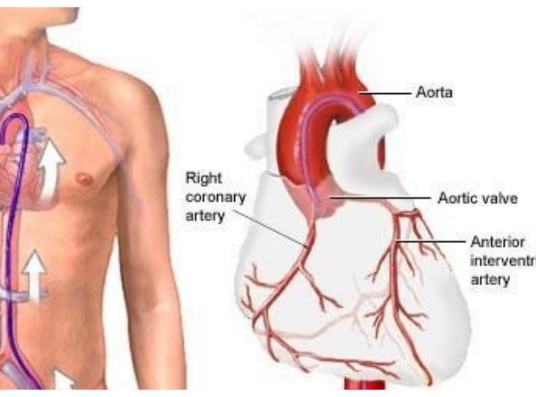
Coronary angiography is a diagnostic procedure performed to check the blood flow and presence of any obstruction (blockages) in coronary arteries that supply blood to the cardiac muscles.

How Coronary Angiography is done?

This procedure is done through the radial artery that is present in the wrist and the femoral artery (thighs). Anyone location is chosen to do this procedure. The sheath is placed within the artery using a needle. A tube-like structure known as the diagnostic catheter is passed through the artery towards the aorta ( the biggest blood vessel arising from the heart). The end of the catheter is placed at the opening of the arteries and a contrast dye is injected into the coronaries.

At the same time, fluoroscopy is done which permits the visualization of the flow of the dye through the coronaries and can be seen on a screen. Fluoroscopy uses X-rays to visualize the coronaries. Images are then acquired at different angles to get a complete idea of the coronaries. The catheter is removed along with the sheath from the artery. The wound is then dressed and heals with proper post-care.

Is it painful? Does it create a wound?

A small puncture hole on the skin about 2 mm in diameter is created using a needle puncture. It is done under local anesthesia and does not require any stitches. When properly done it is painless and all that is felt is a tiny prick of the needle.

Does it require admission to the hospital?

It can be done as an out-procedure and the patient can go home on the same day itself. Usually, patients can be discharged after 4 hrs when done via the radial artery and 6 hours when done via the femoral artery.

Is it a dangerous or life-threatening procedure?

It is a relatively safe procedure and needs to be done only by an expert. As it is an invasive procedure, complications may arise depending on various factors involved in a patient’s medical history. Some minor complications that may arise include hematoma at the puncture site, vasospasm, minor allergic reaction to the dye. Very rarely, life-threatening complications like myocardial infarction, arrhythmias, anaphylaxis, stroke, renal failure, or death may occur. However, these are extremely rare cases and occur in less than 1% of patients.

Can coronary angioplasty be done immediately after angiography?

Yes. Angioplasty which involves stenting the blockages seen on angiography can be performed immediately after angiography depending on the clinical circumstances.

Check out our recent blog – https://www.saideephospital.com/2020/11/29/vision-impairment-cases-rise-during-the-pandemic/

Stay tuned for regular heath updates – https://www.instagram.com/saideephealthcareofficial/

Kangaroo Baby Care

Kangaroo Care is a practice of holding a baby that involves skin-to-skin contact. The baby, who is usually naked apart from a diaper, is placed in an upright position against a parent’s bare chest. Both mothers and fathers can do kangaroo care. It’s often used with premature infants while they’re still within the hospital.

What is kangaroo care?

Holding your baby close to your chest is a surreal experience that can help build the bond between you and your new family member. This type of touch isn’t just for bonding — it’s also medically beneficial for your infant. Kangaroo care is a method of holding your baby close to your chest. It allows for skin-to-skin contact between you and the baby. The process involves that during each session, your baby will be placed (naked except for a diaper and hat) on your chest (also bared to allow skin-to-skin) for up to a few hours. A blanket, shirt, hospital gown, or robe will be wrapped around you and over your baby’s back for warmth. This wrapping of your infant into your chest looks very much like a mother kangaroo holding her baby in her pouch — which is where the name kangaroo care comes from.

kangaroo care

“Kangaroo care first started in the 1970s, as a means to promote bonding and early breastfeeding in full-term infants,” is believed. “In the late 70s, this practice was extended to preterm infants due to over-crowded nurseries, high mortality rates, high infection rates and a lack of resources, like warming devices, known as isolettes. Fast forward nearly 50 years, and the practice of kangaroo care is frequently used in NICUs around the world, due to its profound benefits to both mother and infant.”

We understand that the concept of skin-to-skin is much like kangaroo care, and in many cases, the terms are used interchangeably. In the current day, skin-to-skin is typically a term used for full-term infants, describing how much of the first hours and days of the infant’s life are spent against the mother’s chest, promoting both bonding and breast milk production. Kangaroo care is more often used when referring to the care of a pre-term baby in the NICU receives.

What are the benefits of kangaroo care?

There are many benefits of kangaroo care. It’s not only good for both premature and full-term babies but also for the parents. Both the mother and the father can practice skin-to-skin bonding with the baby.

The benefits of kangaroo care to your baby include:

  • Stabilizing your baby’s heart rate.
  • Improving your baby’s breathing pattern and making the breathing more regular.
  • Improving oxygen saturation levels (this is a sign of how well oxygen is being delivered to all of the infant’s organs and tissues).
  • Gaining in sleep time.
  • Experiencing more rapid weight gain.
  • Decreasing crying.
  • Having more successful breastfeeding episodes.
  • Having an earlier hospital discharge.

The benefits of kangaroo baby care.

  • Improving bonding with your baby and the feeling of closeness.
  • Increasing your breast milk supply.
  • Increasing your confidence in the ability to care for your new baby.
  • Increasing your confidence that your baby is well cared for.
  • Increasing your sense of control.

Why does kangaroo care work?

The benefits of kangaroo baby care listed above have all been demonstrated in research studies. In fact, studies have found that holding your baby skin-to-skin, it can stabilize the heart and respiratory (breathing) rates, improve oxygen saturation rates, better regulate an infant’s blood heat and conserve a baby’s calories.

When a mother is practicing kangaroo care, her infant typically snuggles into her breasts and falls asleep within a couple of minutes. The breasts themselves are shown to vary in temperature to match your baby’s temperature needs. In other words, your breasts can increase in temperature when your baby’s body is cool and may decrease in temperature when the baby is warm.

The extra sleep that your infant gets while snuggling with mom and therefore the assistance in regulating blood heat helps your baby conserve energy and redirect calorie expenditures (use) toward growth and weight gain. Being positioned on mom also helps to stabilize your infant’s respiratory and heart rates. Research has also shown that practicing kangaroo care can have a positive impact on your baby’s brain development.

Do check out our recent blog – https://www.saideephospital.com/2020/12/12/breast-lumps-may-or-may-not-be-malignant-but-they-should-not-be-ignored/

Stay tuned for regular updates – https://www.instagram.com/saideephealthcareofficial/

Breast lumps

Breast lumps may or may not be malignant, but they should not be ignored.

Breast lumps are a common occurrence in women. While only 10 – 20% of breast lumps are malignant, the majority are benign lesions such as fibroadenoma in teenagers and young adults, and fibrocystic change and cysts in older women.

Only approximately 10 to 20% of breast lumps are malignant. You might be surprised to find a breast lump, but it’s important to remember that it may not affect your long-term health. Early detection of breast cancer can save lives, and women aged 40 and above are strongly encouraged to undergo regular mammographic screening.

Breast lumps

How Your Breasts Should Feel

Breast tissue varies inconsistently, with the upper-outer part of your breast being firm and the inner-lower parts feeling somewhat softer. If you are a woman, your breasts can become more tender or lumpy during your menstrual cycle. Breasts tend to get less dense as you get older.

It is important to be familiar with how your breasts normally feel so you are aware of changes. We do not recommend to examine this on your own because there’s little evidence that breast self-examination reduces the risk of dying from breast cancer.

In fact, breast self-examination may cause harm because you’re more likely to find a noncancerous lump, which could be a source of worry. In some cases, this may lead to unnecessary medical procedures to ensure that the lump is benign.

Instead of performing breast self-examination, most experts recommend that women simply be aware of what their breasts normally look and feel like. You should report any changes or concerns to your doctor.

  • a lump changes or grows larger
  • your breast is bruised for no apparent reason

Signs You Should See a Doctor

Remember, most breast lumps are noncancerous. However, you should make an appointment to see your doctor if:

  • you discover a new lump
  • an area of your breast is noticeably different from the rest
  • a lump does not go away after menstruation
  • the skin of your breast is red or begins to pucker like an orange peel
  • you have an inverted nipple (if it was not always inverted)
  • you notice a bloody discharge from the nipple

At Saideep hospital, we recognize the needs women have when it comes to selecting and receiving expert consultations and treatments for any type of breast disease, cancerous, and non-cancerous. We offer a one-stop solutions center, dedicated to looking after breast health.

Our breast imaging center enables women to have their breast ultrasonography, surgical consultation, minimally invasive biopsies, and procedures all under one roof, and with quick turnaround time.

The center is governed and dictated by a multi-disciplinary team of breast surgeons, radiologists, reconstructive surgeons, oncologist together with pathologists and physiotherapists.

With digital mammography, more cancers are getting detected, earlier. It is also significantly more accurate in detecting breast cancers at the earliest stage, or when it is hidden by dense tissues. A screening mammogram is generally recommended for women age above 40 years of age or earlier if she has a family history of breast cancer.

We can help you if you

  • Discover a breast lump
  • Are concerned about your breast cancer risk
  • Have an abnormal mammogram and/or breast ultrasound report
  • Have an abnormal breast biopsy result
  • Have been diagnosed with breast cancer and seek treatment
  • Want a second opinion
  • Need arm physiotherapy for prevention or management of shoulder stiffness, reduced arm mobility and/or lymphedema
  • Want to learn about breast self-examination

Our Range of Services

  • Breast imaging studies such as mammography and ultrasound
  • Image-guided biopsy of breast lesions
  • Minimally invasive breast surgery including core biopsy or lumpectomy
  • Oncologic breast surgery including oncoplastic surgery and breast reconstruction where necessary
  • Adjuvant chemotherapy and radiotherapy including neoadjuvant chemotherapy for downstaging of breast cancers
  • Breast self-examination training and counseling
  • Emotional and psychological support by medical professionals

Check out our recent blog – https://www.saideephospital.com/2020/12/10/delayed-cord-clamping-in-preterm-deliveries-the-benefits-of-dcc/

Stay tuned for daily health updates – https://www.instagram.com/saideephealthcareofficial/

Hi, How Can We Help You?